Major Diseases
The Cardiovascular Center is taken care patients with the following diseases.
-
Valvular Disease
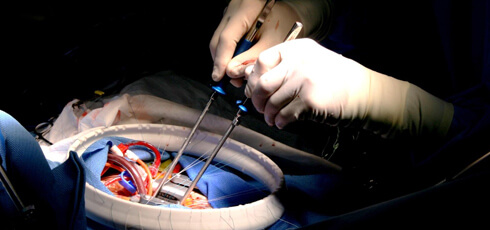
The characteristics of this disease are abnormalities in the cardiac valve that cause palpitations on exertion and breathing difficulties (cardiac failure). We conduct operations, mainly repair the valves but in some cases replace the valve with artificial one.

-
Adult Congenital Heart Disease
We are the first facility in Japan where cardiovascular specialists treat and follow-up adults with congenital heart diseases.

-
Aortic Disease
We repair or replace the aorta, the largest blood vessel in the body.
-
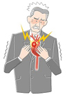
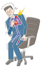
Ischemic Heart Disease
The characteristic of this cardiovascular disease is chest pain and shortness of breath. We provide current and updated therapy.

-
Arrhythmia
We make a diagnosis and proper treatment by thoroughly explaining the various types of irregular pulses, their symptoms and prognosis.
-
Cardiac Failure
Thorough examinations and treatment are necessary if you are told that you may have heart failure. We will provide proper advice and management for cardiac failure.
-
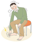
Peripheral Vascular Disease
Peripheral vascular disease is a disorder of the blood vessels in the extremities, arms and legs. The symptoms of this disease are pain and lassitude when walking or holding something heavy.
-
Pulmonary Embolism
We will manage pulmonary embolism, also known as Economy Class Syndrome, and deep vein thrombosis in emergency basis.
-
Atrial Septal Defect
The percutaneous catheter occlusion of Atrial Septal Defect has become available at our hospital. A catheter is inserted from a femoral vein to seal the hole between both atriums, without surgically opening the chest.
-
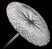
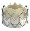
Aortic Valve Stenosis
The treatment for Aortic Valve Stenosis using catheters has become possible. Deformed valves can be fixed or artificial valves can be placed by inserting catheters from the femoral artery or under the subclavian artery, without surgically opening the chest.
Various Currently Available Examination modalities
We offer the best examinations with the newest examination modalities, and apply proper modalities for patients. The following are some of the main modalities for examinations conducted in the Cardiovascular Center.
Echocardiography (Ultrasound)
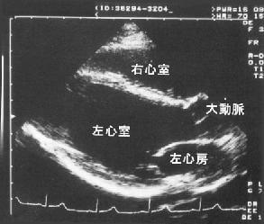
The anatomy and movement of the heart, and conditions of the valves are visualized using echocardiography on the chest or sometimes through esophagus.
Holter Electrocardiography

This examination detects abnormal rhythms of the heart that do not appear in a regular resting 12 leads. This ECG monitors and records the heart beats for 24 hours. We also examine 24 hours blood pressure monitoring at the same time.
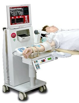
Stress Electrocardiogram
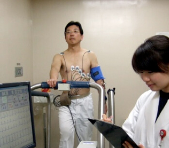
By using treadmills or ergometer exercise machines (exercise bikes), this test examines the changes in electrocardiograms, heart rate and blood pressure when the heart is under stress or loaded condition.
Ankle Brachial Index
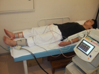
ABI measures the blood pressure of both hands and feet at the same time to measure blood flow velocity and find whether there is arterial sclerosis or not. It also evaluates arterial stenosis in major arteries of extremities and arterial sclerosis.
Vascular Endothelial Function (Flow Mediated Dilation)
This examination evaluates Vascular Endothelial Function by measuring the change in blood vessel width before and after pressure is applied. Vascular Endothelial Function test is sensitive marker for atherosclerosis and stiffness of arteries.
Nuclear Cardiology Examination (Scintigraphy)
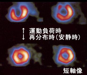
By using the isotopes taken into the cardiac muscle, this examination detects myocardial blood flow, metabolism that is basic factor for the heart and etc.
320-row Multi-slice CT
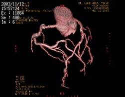
This examination shows anatomy of the whole body including the heart and great vessels. The implementation of the multi-slice CT scan has allowed clear images of the heart and great vessels in 3D fashion , shortened the time of filming and has allowed to clearly visualization of the coronary arteries non-invasively.
MRI Examination
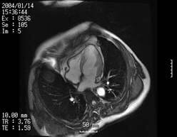
By applying this to the heart and great vessels, it reveals anatomy, function and tissue characterization of the whole body including the heart and great vessels without ionizing radiation. This modality is useful for detecting the disorder of the heart and vessels in various disorders in timely fashion.
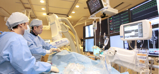
Cardiac Catheterization
This examination observes blood pressure in the heart and vessels, the cardiac and valve function using contrast medium, including coronary arteries. This examination is performed during hospitalization. This examination is one of the best diagnosis methods for cardiac diseases. Also, by using contrast for the aorta and lower limb peripheral artery, the site of stenosis, congenital abnormalities of the heart can be visualized and an accurate diagnosis is possible.

A normal coronary artery
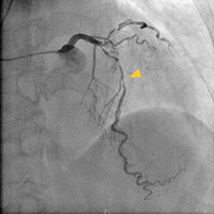
Coronary artery stenosis