
Overview
Since our department’s establishment, we have practiced team medicine. We use state-of-the-art diagnosis and operation equipment including CT/MRI, various Brain Physiology Exams and Nuclear Medicine Scan, Operation Microscopes, Surgery Navigation System, Brain Endoscopy and Surgical lasers. We seamlessly work together with the Emergency Care Center and operate on an on-call system after normal hours to work quickly and effectively.
Diseases and Expertise
Diseases treated at the Neurosurgery Department
| 1. Brain Tumor | 2. Cerebral Vascular Disease | 3. Head Trauma |
|---|---|---|
| Glioblastoma Meningioma Acoustic Schwannoma Pituitary Adenoma Craniopharyngioma Brain Metastasis Intraventricular Tumor |
Subarachnoid Hemorrhage Cerebral Hemorrhage Cerebral Infarction Transient Ischemic Attack Carotid Artery Stenosis Moyamoya Disease Arteriovenous Malformation Dural Arteriovenous Fistula Carotid-Cavernous Sinus Fistula Dissecting Vertebral Artery Aneurysm |
Skull Fracture Epidural Hematoma Subdural Hematoma Traumatic Intracerebral Hematoma Traumatic Epilepsy Cerebrospinal Leakage Otorrhea/Rhinorrhea |
| 4. Spinal Disease | 5. Congenital Disease |
6. Functional Disease |
| Cervical Disc Herniation Cervical Spinal Canal Stenosis (OPLL) Ossification of the Yellow Ligament Spinal Tumor Spinal Trauma Myelomeningocele |
Hydrocephalus Chiari Malformation Arachnoid Cyst Spinal Dysraphism, Myelomeningocele Tethered Cord Syndrome Craniosynostosis |
Hemifacial Spasm Trigeminal Neuralgia Idiopathic Normal Pressure Hydrocephalus (iNPH) Parkinson's Disease Intractable Pain |
Equipment
Diagnostic Equipment: CT, MRI, various Brain Physiology Exams, Nuclear Medicine Scan and Cerebral Angiography (DSA)
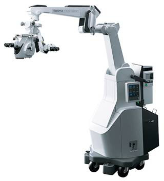
1. Olympus Operative Microscope OME-9000 (ICG・5-ALA Fluorescent Operative Microscope)
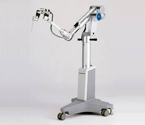
2. Olympus Neurosurgical Endoscope System EndoArm
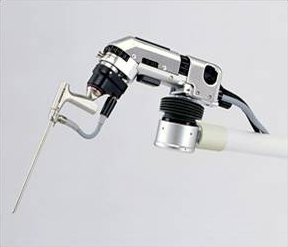
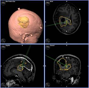
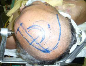
3. Neurosurgical Neuro-navigator

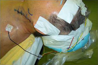
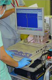
4. Perioperative Neuro electrophysiology monitoring
The neuro electrophysiology monitoring team consisting of professional engineers from the central physiological laboratory conduct perioperative examinations. From here on, brain tumor craniotomy near the speech area will be conducted under neuro electrophysiology monitoring taking waking state operation into consideration.
The neuro electrophysiology monitoring team consisting of professional engineers from the central physiological laboratory conduct perioperative examinations. From here on, brain tumor craniotomy near the speech area will be conducted under neuro electrophysiology monitoring taking waking state operation into consideration.
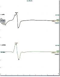
SEP Monitoring

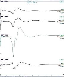
During the brain tumor craniotomy, SSEP is measured and the central channel is identified.
During carotid endarterectomy, the SSEP is measured and cerebral ischemia condition is evaluated.
VEP Monitoring
VEP monitoring is conducted for surgeries relating to the visual pathway including optic nerve tumor and visual center.
![]()

MEP Monitoring
Transcranial stimulation electrodes for MEP during a cervical cord injury surgery

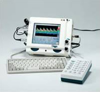
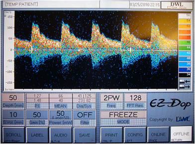
5. GADELIUS Transcranial Doppler Ultrasound Testing Equipment(TCD)
Treatment for angiospasm is mainly conducted; the cerebral blood flow speed is measured during the cerebrovascular spasm period, after subarachnoid hemorrhage.